

The most popular diagnostic imaging method is X-rays (radiographs) or high-quality x-ray scans. The use of diagnostic imaging techniques helps to diagnose an injury or sickness accurately to identify its underlying causes.
These methods in high-quality x-ray scans include computed tomography (CT) scans, MRIs, and x-rays. An x-ray machine fires an x-ray beam through the body to create a picture. Due to the abundance of calcium in bones, the beam does not penetrate bone tissue. The film is then affected by the passing beam, creating an image of the bones.
Benefits Of Using High-Quality X-Ray Scans
The development of CT and the discovery of high-quality x-ray scans marked significant medical developments. X-ray imaging tests are an important medical tool for various operations and examinations. They’re accustomed to non-invasively and painlessly assisting in illness diagnosis and therapy monitoring; supporting the design of medical and surgical treatments; and directing medical staff while they place catheters, stents, or other devices within the body, treat tumors or remove blood clots or other obstructions.
Achieving Perfection in X-Ray Scans: The Quality Process
- An X-ray can be taken in the radiology department of a hospital, a dentist’s office, or a clinic that focuses on diagnostic procedures.
- Once you’re ready, your radiologist or X-ray technician will instruct you on positioning your body for crystal-clear images.
- They might ask you to sit, stand, or lie during the test. They might photograph you with X-ray film or sensors in front of a specific plate.
- They might also ask you to sit or lie down on a specific plate while they move a big camera attached to a steel arm across your body to take X-ray pictures.
- It’s crucial to maintain your stillness while the pictures are being taken.

The Many Uses of X-Ray Scans: Why They’re So Important
Checking The Conditions of The Urinary Tract
Examining the body’s organs for problems is another frequent application for x-rays. Before taking the x-ray, a contrast material is injected into the body as part of an IVP procedure or intravenous pyelogram to help highlight bladder, ureters, and kidney problems. The contrasting fabric makes anomalies in the x-ray images stand out more clearly.
Checking The Mammography
Another frequently used high-quality x-ray scans procedure is mammography, in which patients who want to be checked for abnormal breast growths are requested. X-rays are used in mammograms to provide images of the breast tissue. It is used to identify abnormal growths to assess the need for additional testing.
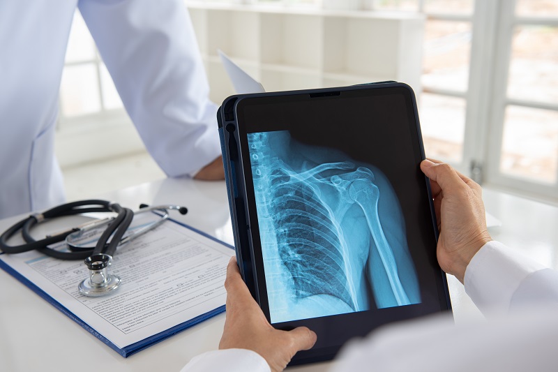
Checking The Bruised bones
Typically, patients who suffer a bone injury are required to have x-rays. An x-ray can still reveal important details about a fracture even if it is used to diagnose it. A doctor can determine from the photographs whether the fracture is full (the bone has split in two) or partial. The x-ray can show whether a bone has moved from its original position.
Conclusion
High-quality x-ray scans, the first type of medical imaging, are still in high demand. It uses electromagnetic waves to create images of the inside organs of bodies. An x-ray provides doctors with black-and-white pictures of your inside structures. Typically, when a doctor needs medical imaging to make a diagnosis, X-rays are the first thing they ask for. X-rays are used not only to make a diagnosis but also to aid in treatment, monitor the development of a condition or damage, and evaluate results after treatment.
No matter because your doctor ordered an x-ray, you can be sure it was a wise choice because it can swiftly and accurately diagnose any illness you may have. X-ray scans are accessible right away, which facilitates finding solutions.


